Design Prototype
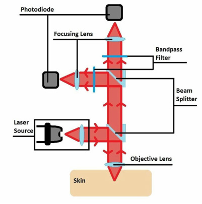
The bloodless glucose monitor measures blood glucose levels using the Raman signal of glucose and an internal standard, hemoglobin. The 785 nm coherent excitation source initiates from the laser source and is collimated to 9 mm in diameter and diverted towards the objective lens by a beam splitter. The objective lens then focuses the collimated light to a near point 4 mm extending from the objective lens, through the skin, onto blood. The objective then collimates the backscattered Raman signal. The Raman signal is then diverted towards the photodetectors via beam splitters. The band pass filter, en route to the photodetectors, filters out irrelevant wavelengths to preserve only 860 nm and 890 nm wavelengths of the respective characteristic glucose and hemoglobin Raman spectral peaks. The photodetector measures the intensity of these light signals and converts them into current signals. Not shown in the picture, the signal is then amplified and converted to from analog to digital by the data acquisition device. The computation circuit interprets the relayed signal and calculates blood glucose concentration to show on the display. The battery provides portable power for the device. The case holds and protects the components.
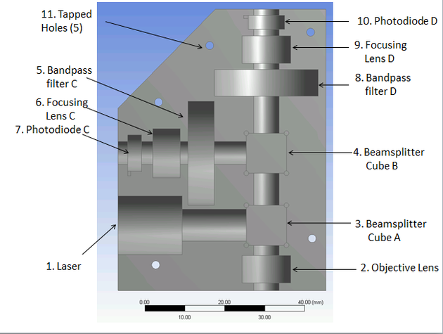
The beam travels from (1) to (3).
It then travels from (3) through (2) and is reflected by the skin placed there back through (2).
It travels back through (3) to (4) where it is split into two beams onto two paths.
The beams travel through (5-6-7) and through (8-9-10).
It then travels from (3) through (2) and is reflected by the skin placed there back through (2).
It travels back through (3) to (4) where it is split into two beams onto two paths.
The beams travel through (5-6-7) and through (8-9-10).
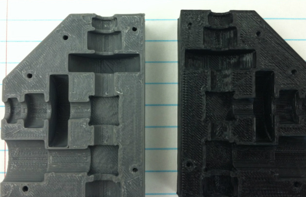
Pictured are the two halves of the designed casing for the optical components.
